Convenient Locations
- Shreveport
- Protected: Texarkana

Diagnostics
Optical Coherence Tomography
The Optical Coherence Tomography, known as the OCT, uses light waves to take a cross sectional scan to provide a detailed image of each individual layer of the retina. This allows the doctor to diagnose conditions such as Vitreomacular Traction, Epiretinal Membrane, Macular Holes, Cystoid and Diabetic Macular Edema, Macular Degeneration, Central Serous Chorioretinopathy and many more pathologies.
Fundus Photography
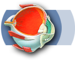
Fundus Photography is an image taken of the internal structures of the eye to include the Retina, Macula, Fovea and Optic Nerve. The patient’s eye is dilated and the photograph is taken. This provides a digital snap shot of the inside of the eye which can be referred back to in the future.
Ultrasonography
B-Scan Ultrasonography provides a noninvasive mean to evaluate and visualize ocular structures which are otherwise impossible to view due to opaque ocular media. This is achieved by the use of high frequency sound waves transmitted to the eye by a diagnostic probe. These sound waves are reflected back from the intraocular surfaces they land upon as an electric signal. This electric signal is transmitted from the diagnostic probe to a monitor to provide an image of the internal structures of the eye. This technique can also be useful in the measurement of Intraocular Tumors, differentiating Optic Nerve Head Drusen from Papilledema, and viewing Ciliary Body Detachments.
Fluorescein Angiography
Flourescein Angiograpy is a procedure which highlights the Retinal and Choroidal circulation. Flourescein Dye is injected into the arm and a camara is then used to image the flow of the dye through the vessels of your Retina and Choroid.
Intravitreal Drug Therapy
Intravitreal Drug Therapy involves the injection of medication into the vitreous humor of your eye.
VEGF Inhibitors
VEGF stands for Vascular Endothelial Growth Factor. VEGFs promote the growth of abnormal blood vessels. VEGF Inhibitors block the growth of these abnormal vessels.
Avastin – (Bevacizumab)
- Avastin is approved by the FDA to treat Colorectal Cancer; studies have shown that Avastin can also effectively treat retinal diseases by inhibiting VEGF. Avastin is given every 4 to 6 weeks. For more information regarding Avastin click here.
Lucentis – (Ranibizumab)
- Lucentis is an FDA approved, anti-VEGF agent, used to block abnormal blood vessel growth and leakage. It is injected into the eye to treat Wet Age Related Macular Degeneration (AMD), Diabetic Macular Edema and Macular Edema following a Branch or Central Retinal vein Occlusion. Lucentis is given as 1 injection per month. For more information regarding Lucentis and patient assistance programs click here.
Eylea – (Afilbercept)
- Eylea is FDA approved to treat Wet- Age Related Macular Degeneration and Macular Edema following Central Retinal Vein Occlusion. Eylea is recommended every 8 weeks following 3 initial monthly injections. For more information regarding Eylea and patient assistance programs click here.
Corticosteroids
Kenalog – (Triamcinolone Acetonide)
- Kenalog, also known as Trimacinolone Acetonide, is used to treat Cystoid Macular Edema and Uveitis.
Ozurdex – (Dexamethasone)
- Ozurdex is a Dexamethasone Intravitreal Implant approved by the FDA to treat Macular Edema following a Retinal Vein Occlusion and Posterior Uveitis. Ozurdex is a biodegradable implant which is injected into the vitreous where it slowly dissolves and releases the corticosteroid dexamethasone. Ozurdex typically is effective for 3 months. For more information regarding Ozurdex click here.
Laser Treatment
The definition LASER is defined as light amplification by stimulated emission of radiation.
Photocoagulation
- Photocoagulation uses a laser to coagulate tissue. The light emitted from the laser is absorbed as thermal energy by the retinal tissue. This thermal energy (heat) causes necrosis of retinal tissue.
- Photodynamic therapy involves the intravenous injection of a photosensitizing drug, verteporfin, which migrates to particular cells within the retina. A laser is then used to activate this drug to achieve its desired affects without using photocoagulation.

Vitreo-Retinal Surgery
Vitrectomy (PPV)
- The Vitreous is a clear, jelly-like substance that fills the center of the eye; it is located behind the lens and anterior to the retina. During a vitrectomy, the vitreous is removed and may be replaced with a gas or air bubble. If a bubble is placed within the eye, it will be slowly absorbed and replaced by the body’s own fluids. In severe cases, the vitreous may be replaced with silicone oil; this oil usually will require removal at a later date. A vitrectomy can be used to treat retinal detachments, macular pucker, macular hole, complications of diabetic retinopathy, infections inside the eye, and a severe eye injury.
Retinal Detachment Repair
- A retinal detachment repair includes a vitrectomy, drainage of fluid beneath the detachment, air for gas exchange or in more complex situations air for silicone oil exchange, then is completed with laser or cryotherapy to seal the detachment.
Scleral Buckle
- A sclera buckle is a flexible band which is placed around the eye to counteract the force causing a retinal detachment. The fluid underneath the detached retina will be drained to allow the retina to settle back into its normal position.
Vitreo-Retinal Associates—helping you keep sight of what’s important!

 Contact us
Copyright © 2005-2024 Vitreo-Retinal Associates. All Rights Reserved